Blood Vessels Labeled Simple - Pin on Anatomy and Physiology Models / August 17, 2020 so, you want to learn.. While most blood vessels are located deep from the surface and. Veins return blood back toward the heart. Thus, rvdm provides an accurate quantification of blood flow velocity, and due to its compatibility with simple sxyt imaging, allows blood flow measurement. Arterioles connect with even smaller blood vessels called capillaries. Subscribe to the operation ouch official channel!
Veins return blood back toward the heart. Veins (in blue) are the blood vessels that return blood to the heart. Molly smith dipcnm, mbant • reviewer: Transcribed image text from this question. Terms in this set (6).
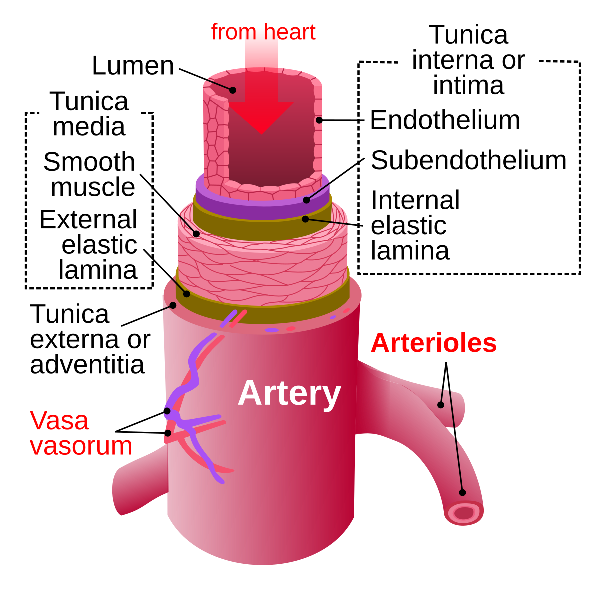
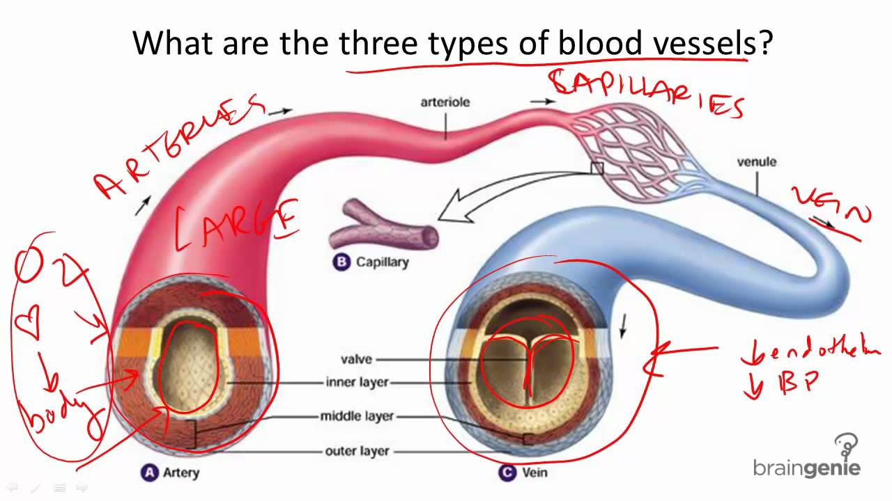
Artery - Wikipedia from upload.wikimedia.org Blood vessels labeled simple : • identification of blood vessels as arteries, capillaries or veins from the structure of their walls. Blood travels from the heart in arteries, which branch into smaller and smaller vessels, eventually becoming arterioles. Related posts of the human blood vessels labeled digestive system free online quiz blood vessel labeling there are five main types of blood vessels: Arterioles, capillaries, venules, are known to respond differently to various stimuli7,9,11. Arterioles connect with even smaller blood vessels called capillaries. The difference in the structural characteristics of arteries, capillaries and veins is attributable to their respective functions. The iliac, femoral, popliteal and tibial (calf) veins are the deep veins in the legs.
Blood vessels are vital for the body and play a key role in diabetes helping to transport glucose and insulin.
This page provides histology support information for blood vessel structure. Retinal_blood_vessel_segmentation.ipynb this is a simple implementation neural net with. All blood vessels are specifically structured to perform their function. Blood vessels 2 labeled palmar arch digital artery right femoral a right femoral v great saphenous vein left popliteal a right anterior tibial a. Through the thin walls of the capillaries, oxygen and nutrients pass from blood into tissues, and waste products. The best websites voted by users. Although whole blood does not leave the vessels, components of the plasma and tissue fluids can be exchanged through the walls of the tiniest vessels, the capillaries. Carry blood towards the heart (usually deoxygenated blood, except for the pulmonary vein). Allows diffusion of gases and nutrients from blood into the body cells. Related posts of the human blood vessels labeled digestive system free online quiz blood vessel labeling there are five main types of blood vessels: Transcribed image text from this question. The inner lining is the endothelium and is surrounded by subendothelial connective tissue. Simple squamous epitheliumendothelium in blood vessels blood vessel color images
It is made up of. The capillaries also connect the branches of arteries and to. Between arteries and veins, there is a network of. ⇒ click on the diagram to show / hide labels. • identification of blood vessels as arteries, capillaries or veins from the structure of their walls.
8.7.2 Blood Vessel Structure and Function - YouTube from i1.ytimg.com Arterioles connect with even smaller blood vessels called capillaries. Hma practical 3 for monday july 23 and wednesday july 25. While most blood vessels are located deep from the surface and. This page provides histology support information for blood vessel structure. The inner lining is the endothelium and is surrounded by subendothelial connective tissue. ⇒ click on the diagram to show / hide labels. Blood vessels are intricate networks of hollow tubes that transport blood throughout the entire body. The capillaries also connect the branches of arteries and to.
It is made up of.
Blood travels from the heart in arteries, which branch into smaller and smaller vessels, eventually becoming arterioles. Although whole blood does not leave the vessels, components of the plasma and tissue fluids can be exchanged through the walls of the tiniest vessels, the capillaries. Blood vessels are intricate networks of hollow tubes that transport blood throughout the entire body. These vessels transport blood cells, nutrients, and oxygen to the tissues of the body. Related posts of the human blood vessels labeled digestive system free online quiz blood vessel labeling there are five main types of blood vessels: Deep veins, located in the center of the leg near the leg bones, are enclosed by muscle. Capillaries surround body cells and tissues to deliver and absorb oxygen, nutrients, and other substances. By printing out this quiz and taking it with pen and paper creates for a good. The best websites voted by users. This page is about blood vessel histology slide labeled,contains artery microscope slide. Hma practical 3 virtual slides. It is made up of. Differentiate among the structure of arteries, veins, and capillaries.
They also take waste and carbon dioxide away from the tissues. Molly smith dipcnm, mbant • reviewer: Terms in this set (6). ⇒ click on the diagram to show / hide labels. These vessels transport blood cells, nutrients, and oxygen to the tissues of the body.
Pin on Histology - Vascular from i.pinimg.com The blood vessels are the components of the circulatory system that transport blood throughout the human body. It is made up of. ⇒ click on the diagram to show / hide labels. Through the thin walls of the capillaries, oxygen and nutrients pass from blood into tissues, and waste products. Hma practical 3 virtual slides. These vessels transport blood cells, nutrients, and oxygen to the tissues of the body. Although whole blood does not leave the vessels, components of the plasma and tissue fluids can be exchanged through the walls of the tiniest vessels, the capillaries. • identification of blood vessels as arteries, capillaries or veins from the structure of their walls.
Between arteries and veins, there is a network of.
The best websites voted by users. Does not form part of the actual practical class based upon the virtual slides. All blood vessels are specifically structured to perform their function. They also take waste and carbon dioxide away from the tissues. August 17, 2020 so, you want to learn. Vessels that carry blood away from heart, surrounded by thick layer of smooth muscle, high levels of bp review: Blood vessels cannot function properly when inhibited by vascular diseases. The blood vessels, together with the four chambers of the heart, form a closed system in which blood is carried to and from the tissues. Veins return blood back toward the heart. Blood vessels can be damaged by the effects of high blood glucose levels and this can in turn cause damage to organs, such as the heart and eyes, if significant blood vessel damage is sustained. To print or download this file, click the link below Between arteries and veins, there is a network of. Blood vessels labeled simple :
Blood travels from the heart in arteries, which branch into smaller and smaller vessels, eventually becoming arterioles blood vessels labeled. The intima is a simple epithelium made up of a single layer of flat epithelial cells.


0 Komentar